Label-free optical nanoscopy, free from photobleaching and photochemical toxicity of fluorescence labels and yielding 3D morphological resolution of <50 nm, is the future of live cell imaging. 3D-nanoMorph breaks the diffraction barrier and shifts the paradigm in label-free nanoscopy, providing isotropic 3D resolution of <50 nm. To achieve this, 3D-nanoMorph performs non-linear inverse scattering for the first time in nanoscopy and decodes scattering between sub-cellular structures (organelles).

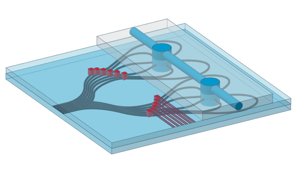
3D-nanoMorph innovatively devises complementary roles of light measurement system and computational nanoscopy algorithm. A novel illumination system and a novel light collection system together enable measurement of only the most relevant intensity component and create a fresh perspective about label-free measurements. A new computational nanoscopy approach employs non-linear inverse scattering. Harnessing non-linear inverse scattering for resolution enhancement in nanoscopy opens new possibilities in label-free 3D nanoscopy.
3D-nanoMorph will be applied to study organelle degradation (autophagy) in live cancer cells over extended duration with high spatial and temporal resolution, presently limited by the lack of high-resolution label-free 3D morphological nanoscopy. Successful 3D mapping of nanoscale biological process of autophagy will open new avenues for cancer treatment and showcase 3D-nanoMorph for wider applications.
Krishna Agarwal’s expertise of 14 years spanning inverse problems, electromagnetism, optical microscopy, integrated optics and live cell nanoscopy paves path for successful implementation of 3D-nanoMorph.